Detergent
Extraction
200µl of each brain homogenate was extracted in an equal volume of
2X lysis buffer (20mM Tris PH 7.4, 1%SDS, 1% Triton X-100, 1%
deoxycholic acid). Insoluble material was removed by centrifugation
at 4000rpm for 5mins at 4°C.
PK
Digestion
For PK digestion 100µl of detergent extracted homogenates were
treated with 50µg/ml proteinase K for 1hr at 37°C. PK digests were
stopped by adding PMSF to a final concentration of 1mM.
PGnase Digestion
For digestion with PGNase F 3µl of denaturation buffer (NEB) was
added to 25µl of PK treated extracts and incubated at 100°C for
10mins. 3µl of water, 4µl NP40 and 4µl of 10X PGNase buffer were
added to denatured extracts and duplicate samples +/- PGNase F
(1µl; 500U/µl were incubated at 37°C for 2hr.
Precipitation of
Digested Protein
Protein was precipitated by adding 4 vols of ice-cold methanol,
incubated at –20°C for 30mins and pelleted by centrifugation for
30mins at 14,000rpm. All traces of supernatant were removed and
pellets were resuspended in 30µl of 1X lysis buffer.
Western Blotting
and Visualisation
An equal volume of 2X denaturation buffer was added and samples
were heated to 100°C for 10 mins. A total of 15µl of sample was
loaded onto a 16% Tris-glycine gel that was run for 60 mins at 100
volts, 120mA. Gels were transferred onto PVDF and blocked for 1hr
in 5% (in PBS)non-fat milk . Blots were incubated o/n at room temp
with 1o antibody (1µg/ml 3F4) in PBS-Tween (0.05%).
Blots were then washed 4x5mins in PBS-Tween and incubated for 1hr
in 2o antibody (peroxidase conjugated goat anti-mouse
1:10,000) in PBS-Tween (0.05%). After 4 washes in PBS the blot was
developed using ECL-plus and visualised by UVP image analysis.
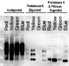
Note: The final loading volumes of the digested protein are
equivalent to 3.2µl of the homogenate. Loadings of undigested
samples were adjusted to represent 3.2µl of homogenate.
Western Blot
| Reference in Figure |
Actual Sample Code |
sample chr$ |
| Red |
NHBX0/0001 |
spCJD(M/M) |
| Green |
NHBX0/0002 |
spCJD(M/M) |
| Blue |
NHBY0/0003 |
nvCJD(M/M) |
| Yellow |
NHBX0/0004 |
spCJD(M/V) |